
WRITTEN BY: Natalie Woolley
BTrngDev, DipVET, DipVN (ECC & Surgical), ISFM AdvCertFB, Cert IV (VN & CGC), Cert IV TAE, MHFA, RVN
PUBLISHED: 30 MARCH 2017
REVIEWED/ UPDATED: 9 OCT 2025
Fluid therapy treatment is one of the backbones of medical therapy in veterinary medicine and a daily task for the veterinary nurse. Its ubiquitous across all modalities of treatment and medical management and a field in which us Veterinary Nurses can truly shine. Here are some tips and cheats to help you flow through fluid monitoring!
Patient assessment
Lets start at the beginning with patient assessment. We are all doing this in our own unique way, but following the same basic steps. Whilst what you do might not change very much, this might help you change how you approach the subjective parts of patient assessment.
Do your skin turgor test in more than one location!
The turgor test, or skin tenting assesses skin elasticity, and this can be different in different locations, and is also affected by other factors outside hydration. Consider things such as age: geriatric patients may have reduced elasticity; paediatric patients may maintain elasticity despite dehydration! Those with a lean body weight may have a prolonged skin tent in the absence of clinical dehydration; whilst fat patients are like paediatric patients where elasticity can be falsely preserved despite dehydration.
We all use the skin over the dorsal scapulae for the skin tent test, but we can also use skin over the thoracic vertebrae, on the lateral scapula and even on the cranium between the ears (Donohue, 2012 pp 17). For patients that tense and resent the dorsal scapulae pinch, this can be useful to remember. Skin turgor should be assessed considering the other parameters checked during an assessment such as mucous membrane colour and moistness and refill, heart rate, pulse quality, blood pressure etc, on its own its not a reliable indicator of hydration status.
Looking at mucous membranes (MM) is a simple, quick task. As vet nurses, we are all confident at doing this. When making your assessment ask yourself these questions: Has the dog been panting (so are the MM tacky from this?); Is there ptyalism, nausea or vomiting present (which makes MM’s moist and slippery). If the patient has been asleep the mucous membranes might be slightly pale. What is the temperature in the room, this can affect peripheral vasodilation and constriction and affect the MM colour.
The subjective data we collect compliments the objective (measurable) information, such as Body weight, PCV, TP, TS, USG to determine the hydration status of the patient. With ongoing monitoring if the same things are assessed at regular intervals, we can build a clear picture of the patients progress. Add these tasks onto your chart at set times. Most measurements don’t cost us anything extra (vitals, blood pressure), but can make a significant difference to your patient.
If your measurable parameters and vitals aren’t changing toward normal, then the fluid therapy plan needs to be adjusted. Your patient might have worsened, or have tachycardia despite adequate analgesia and you recommend to the vet that a bolus infusion of shock rates for 15-30 mins is given and reassess the patient after that. Or you might see that there were less losses (vomiting, diarrhoea) than anticipated, so need to decrease the rate to accommodate this. It’s a dynamic process and needs veterinary nursing attention to detect and reports these subtle differences. These small things add up to massive improvement in patient care and earlier discharge of healthy patients.
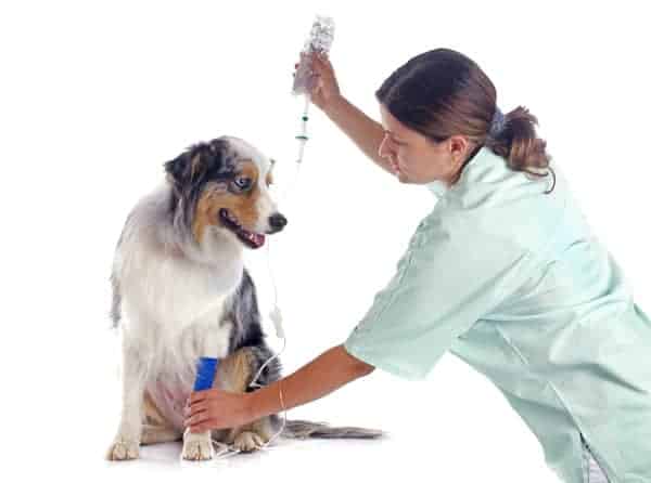
Placing catheters
The technical aspects of starting fluid therapy can be easily done by a pair of Veterinary Nurses assisting each other. Being familiar with the different venepuncture locations can help ensure you have ongoing secure venous access. The cephalic vein, whilst convenient, isn’t always the best site.
Aggressive dog? Think about using a pedal or saphenous vein to avoid the bitey end and reduce stress on the patient. Is the patient nauseous and drooling? Consider avoiding the cephalic in case it gets contaminated and compromised from all that moisture – perhaps use a hind leg vein if you can.
Small patients might have a larger medial saphenous vein than cephalic. Or maybe an intraosseous cannula is a better option in your tiny paediatric cases. Think outside the square, and upskill your team to help improve your fluid therapy care.
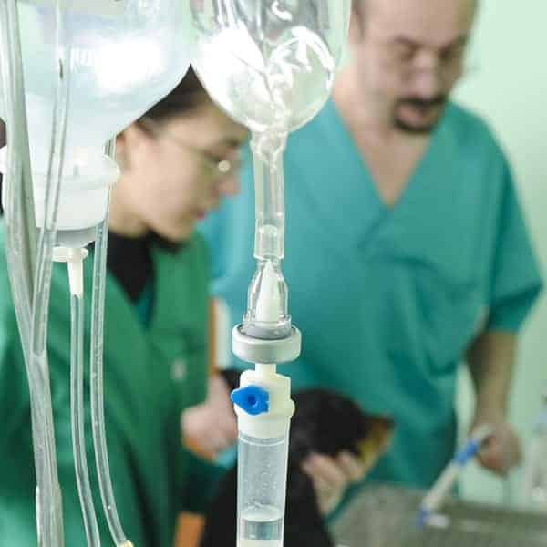
Monitoring fluid therapy plans
 When it comes to charting and monitoring, it can be overwhelming with what to monitor when and how to record it all.
By doing your Fluid Therapy Plan you get a known fluid goals to reach, and know roughly how long it’s going to take you to reach them (depending on patient progress). This helps work out what and when to monitor, and what it means through the day.
Set the Volume to Be Infused (VTBI) goal for the day.
For a simple dehydration, we aim to correct the full dehydration deficit in 12-24 hours. For example, it might be 1840ml in 24 hours for your patient. Delivering this at 76ml/hr will meet your target. (1840 / 24 = 76). If you don’t set this on the infusion pump, at least write it on the chart.
For a combination of dehydration and hypovolaemia, it will be necessary to calculate a bolus volume to deliver over a shorter time period (15-30 minutes) before reassessing perfusion parameters in the patient. Multiple bolus deliveries may be needed before eventually changing to a slower rate to deliver the remaining rehydration volume over a longer period.
Regular monitoring (at least 3-4 times a day, more often for critical patients) will ensure you are meeting your patient’s needs, avoiding fluid overload, going to meet the fluid target(s) and allow you to adjust things when necessary. Some tips to make this easier for you:
- Reset your VI (Volume Infused) to 0 on the pump at the start of each day – this allows you to monitor what has been infused.
- Regularly check that the VI meets what the patient should have received (target volume), though adapt to your patient’s needs – these may change and a new target volume might need to be recalculated. Calculate how much should have been infused and write this on the chart as targets throughout the day.
- VI is a simple calculation, the hours since last checked X by the hourly rate (and add on any previously infused totals).
- Set timers to check your drip pumps at these set times, to see if the VI on each patient matches the target volume at that time
- If not, act to correct it! (Alert vet, replace catheter, change bag, flush catheter, adjust rate if safe to do so)
- Do visual assessment and weigh your patient at regular intervals over the day, such as when toileting, to assist with monitoring. We want to assess perfusion parameters when delivering bolus fluid resuscitation for hypovolaemia (HR, CRT, MM, pulses, BP, extremities and body temperature). When assessing a patient’s response to rehydration fluids, we look for improved skin turgor, moist MM, increased body weight and urine outputs, plus a change in USG.
- Weigh cat litter trays, or absorbent bedding, or before and after urination to determine urine output (if you don’t have a urinary catheter indwelling).
- Remember to check any closed-system wound drains, as the drainage in these is considered a fluid output.
- Remember that ongoing losses should be assessed through inputs and outputs and incorporated into the fluid plan. Using a Fluid Balance Chart (FBC) can assist with monitoring and recording these.
I hope these simple tips help you better assess and monitor your patients, and help you streamline monitoring your patients on fluids. If you want to learn more on fluid therapy, we have a great short course for veterinary nurses.
Happy Veterinary Nursing!
References
- Donohoe, C 2012, Fluid Therapy for Veterinary Technicians and Nurses, Wiley-Blackwell, US.
- Pardo, M., Spencer, E., Odunayo, A., Ramirez, M. L., Rudloff, E., Shafford, H., Weil, A., & Wolff, E. 2024, ‘2024 AAHA Fluid Therapy Guidelines for Dogs and Cats’, Journal of the American Animal Hospital Association, vol. 60, no.4, pp.131–163.
want to learn more?
Fluid therapy
SHORT COURSE
Fluid therapy is an integral part of the modern veterinary clinic. This subject will teach students about the various fluids available, when to use them, and how to monitor patients receiving fluid therapy. Students will also learn about osmosis, and how to calculate correct fluid rates.
Delivered over 13 topics ranging from water and electrolyte balance in the body, to administration of fluids and monitoring your patients, this course will ensure you are well equipped to assist the veterinarians and provide excellent nursing care to all patients receiving fluid therapy.
About the author: View Natalie Woolley expert profile, qualifications and all articles